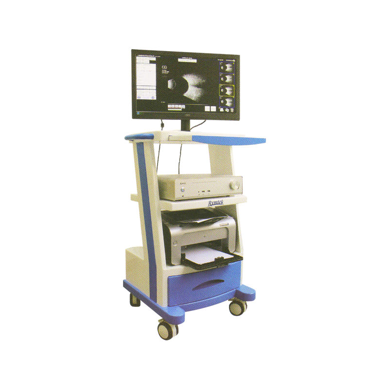
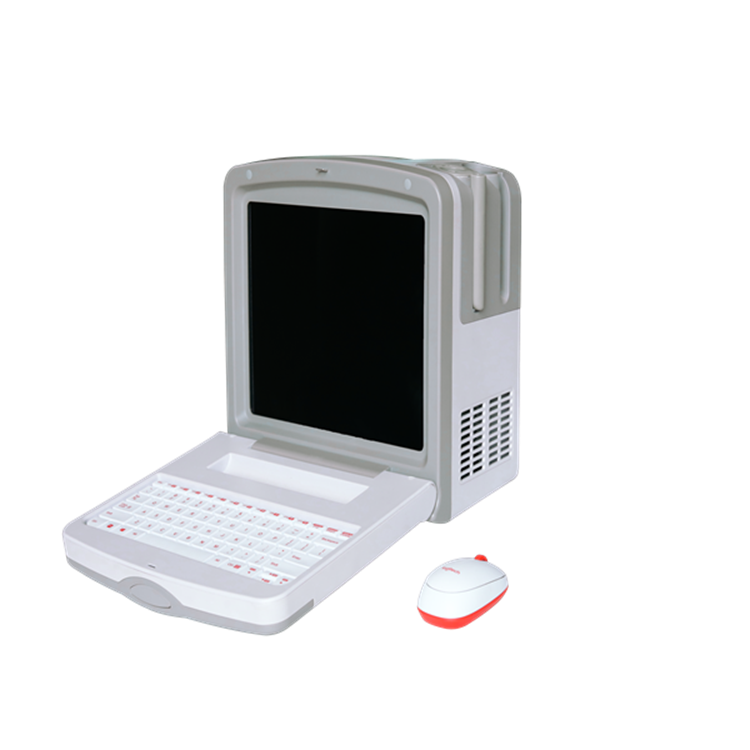
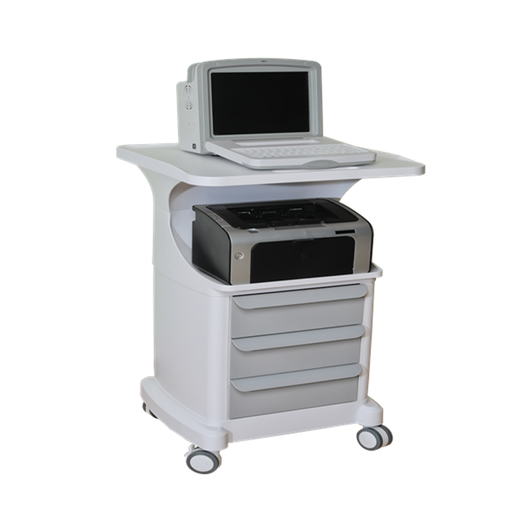
ophthalmic ultrasound scanner


中文介绍
- 定义与用途:眼科超声诊断仪是一种利用超声波技术对眼部疾病进行诊断的医疗设备。它能清晰显示眼部组织结构,为眼科医生提供准确的病情信息,辅助诊断多种眼部疾病,是眼科临床检查的重要工具。
- 可治疗病症(注:超声诊断仪主要用于诊断,并非直接治疗,但可为治疗提供关键依据)
- 眼内异物:可精准确定异物的位置、大小、形状及与周围组织的关系,为异物取出手术提供重要参考,避免手术对眼部组织造成不必要的损伤。
- 视网膜脱离:清晰显示视网膜的形态、脱离范围及程度,帮助医生判断病情严重程度,制定合适的治疗方案,如选择巩膜扣带术、玻璃体切除术等不同术式。
- 玻璃体疾病:包括玻璃体混浊、积血、后脱离等。通过超声图像,医生能了解玻璃体病变的性质和范围,评估病情发展,为治疗提供依据,例如判断是否需要药物治疗或手术干预。
- 眼内肿瘤:可以发现眼内肿瘤的存在,并初步判断肿瘤的位置、大小、边界及内部回声特点,辅助医生鉴别肿瘤的良恶性,为后续治疗(如手术切除、放疗、化疗等)提供关键信息。
- 工作原理:仪器产生高频超声波(一般频率在 5 – 15MHz),通过探头将超声波发射到眼部组织。超声波在眼部不同组织界面会发生反射、折射和散射,反射回来的超声波被探头接收,转换为电信号,经过仪器的信号处理和分析系统,将这些电信号转化为图像信息,以二维或三维的形式在屏幕上呈现出来,医生根据图像特征来诊断眼部疾病。
- 优势
- 无创检查:无需侵入性操作,不会对眼部组织造成创伤,患者接受度高,尤其适用于不能耐受侵入性检查的患者,如儿童、老年人或眼部外伤急性期患者。
- 实时成像:能够实时显示眼部组织结构和病变情况,医生可以在检查过程中动态观察,及时发现病变特征,做出准确判断。
- 不受眼部屈光介质影响:对于角膜混浊、白内障等导致眼部屈光介质不清的情况,超声诊断仪仍能清晰显示眼内结构,这是其他光学检查方法(如眼底镜检查)所无法比拟的优势。
- 操作简便、检查迅速:整个检查过程相对简单快捷,一般几分钟内即可完成,能为急诊患者争取宝贵的诊断时间,也提高了临床工作效率。
英语介绍
- Definition and use:The ophthalmic ultrasonic diagnostic apparatus is a medical device that uses ultrasonic technology to diagnose eye diseases. It can clearly display the tissue structure of the eye, providing accurate disease information for ophthalmologists and assisting in the diagnosis of various eye diseases. It is an important tool for ophthalmic clinical examinations.
- Diseases for diagnosis (Note: The ultrasonic diagnostic apparatus is mainly used for diagnosis, not direct treatment, but provides crucial basis for treatment)
- Intraocular foreign bodies: It can accurately determine the location, size, shape of foreign bodies and their relationship with surrounding tissues, providing important references for foreign body removal surgery and avoiding unnecessary damage to eye tissues during the operation.
- Retinal detachment: It clearly shows the shape, detachment range and degree of the retina, helping doctors judge the severity of the condition and formulate appropriate treatment plans, such as choosing different surgical methods like scleral buckling or vitrectomy.
- Vitreous diseases: These include vitreous opacification, hemorrhage, posterior detachment, etc. Through ultrasonic images, doctors can understand the nature and scope of vitreous lesions, evaluate the disease progression, and provide a basis for treatment. For example, it helps to determine whether drug treatment or surgical intervention is required.
- Intraocular tumors: It can detect the presence of intraocular tumors, and preliminarily judge the location, size, boundary and internal echo characteristics of the tumors. This assists doctors in differentiating the benign and malignant nature of the tumors, providing key information for subsequent treatments (such as surgical resection, radiotherapy, chemotherapy, etc.).
- Working principle:The instrument generates high – frequency ultrasonic waves (usually with a frequency range of 5 – 15MHz). The ultrasonic waves are emitted into the eye tissue through a probe. At the interfaces of different eye tissues, the ultrasonic waves will be reflected, refracted and scattered. The reflected ultrasonic waves are received by the probe and converted into electrical signals. After being processed and analyzed by the instrument’s signal processing and analysis system, these electrical signals are transformed into image information, which is presented in a two – dimensional or three – dimensional form on the screen. Doctors diagnose eye diseases based on the image characteristics.
- Advantages
- Non – invasive examination: It does not require invasive operations and will not cause trauma to eye tissues. Patients have a high acceptance rate, especially suitable for patients who cannot tolerate invasive examinations, such as children, the elderly or patients in the acute stage of eye trauma.
- Real – time imaging: It can display the tissue structure and lesions of the eye in real – time. Doctors can dynamically observe during the examination, promptly discover the characteristics of lesions and make accurate judgments.
- Unaffected by the refractive media of the eye: For cases where the refractive media of the eye are unclear, such as corneal opacity and cataract, the ultrasonic diagnostic apparatus can still clearly display the intraocular structure, which is an advantage that other optical examination methods (such as funduscopy) cannot match.
- Simple operation and rapid examination: The entire examination process is relatively simple and fast. Generally, it can be completed within a few minutes, which can save valuable diagnostic time for emergency patients and improve clinical work efficiency.
