fundus angiograph


英文介绍
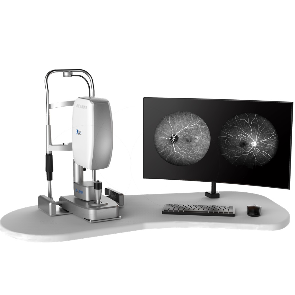
A fundus angiograph is a specialized medical device mainly used in ophthalmology for the diagnosis of fundus diseases. Here is a detailed introduction:
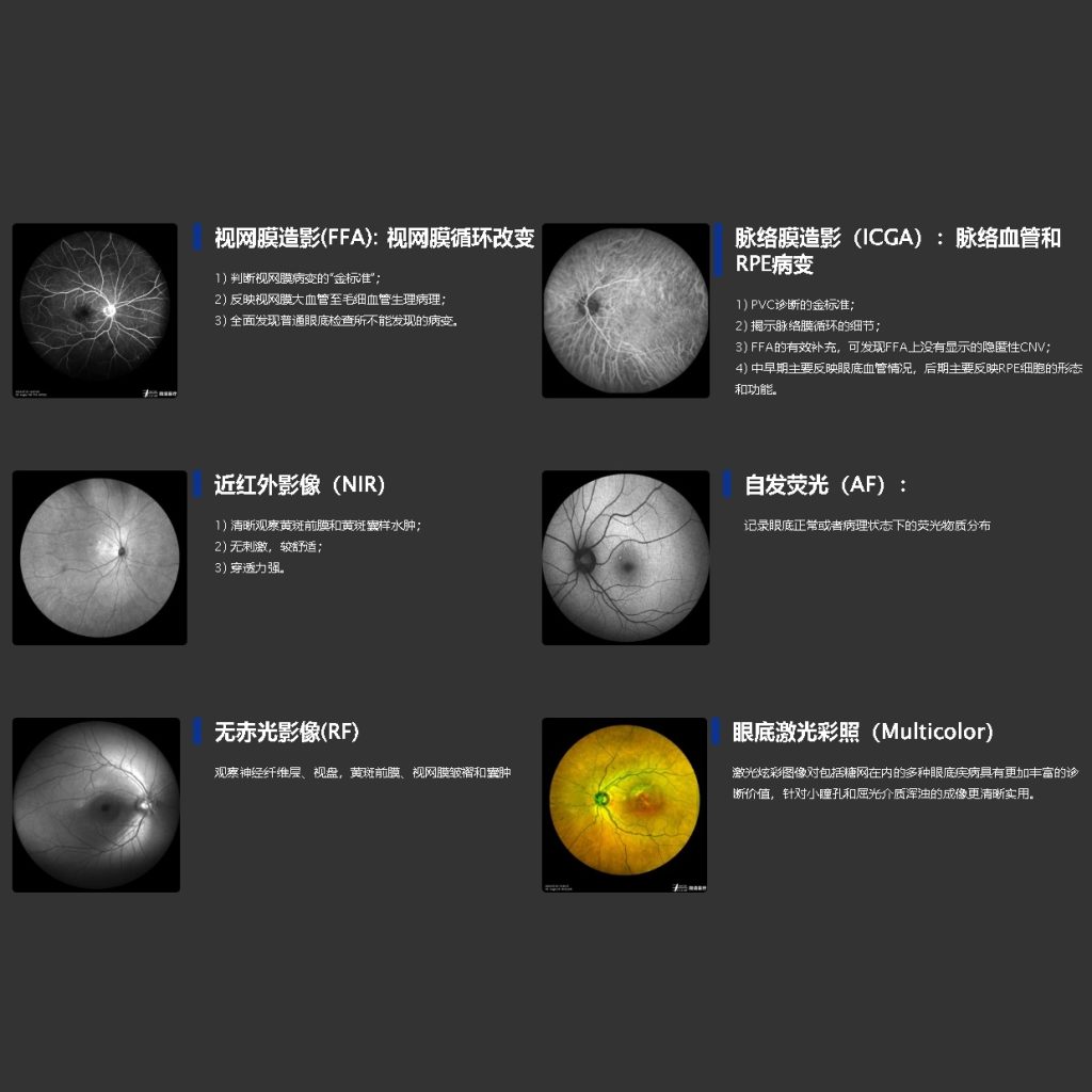
- Working Principle: The fundus angiograph works in conjunction with a contrast agent. Usually, a fluorescent contrast agent like sodium fluorescein is injected intravenously. The contrast agent flows with the blood to the fundus blood vessels. The machine is equipped with special filter plates, which can capture the fluorescence emitted by the contrast agent as it spreads in the fundus blood vessels, so as to visualize the blood – vessel condition of the fundus.
- Structural Composition: It generally consists of an optical system, a lighting system, a camera system, and a control system. The optical system is responsible for focusing the fundus image, the lighting system provides appropriate light sources, the camera system is used to take pictures of the fundus, and the control system is used to adjust parameters such as exposure and shooting time, and to control the injection of contrast agent in some cases.
- Clinical Applications: Fundus angiographs play a crucial role in the diagnosis of various fundus vascular diseases, such as diabetic retinopathy, retinal vein occlusion, and choroidal neovascularization. By observing the dynamic process of contrast agent in fundus blood vessels, doctors can clearly identify the location, extent and nature of vascular lesions, which provides important basis for formulating treatment plans, including guiding fundus laser treatment and surgical interventions.
中文介绍
眼底造影机是一种专门用于眼科领域,以辅助诊断眼底疾病的医疗设备。具体介绍如下:
- 工作原理:眼底造影机需与造影剂配合使用。通常是将荧光素钠等荧光造影剂通过静脉注射的方式注入人体,造影剂随血液流经眼底血管。机器配备特殊的滤光板,能够捕捉造影剂在眼底血管内扩散时所发出的荧光,从而使眼底血管状况得以显像。
- 结构组成:它一般由光学系统、照明系统、摄像系统和控制系统组成。光学系统负责对眼底图像进行聚焦,照明系统提供合适的光源,摄像系统用于拍摄眼底情况,控制系统则用于调节曝光、拍摄时间等参数,部分还可控制造影剂的注射。
- 临床应用:眼底造影机在多种眼底血管性疾病的诊断中起着关键作用,如糖尿病视网膜病变、视网膜静脉阻塞、脉络膜新生血管等。通过观察造影剂在眼底血管内的动态过程,医生能够清晰地识别血管病变的位置、范围及性质,为制定治疗方案提供重要依据,包括指导眼底激光治疗以及手术干预等。
